
Microbiology @ IHS Laboratory (BioLab).
BioLab Office

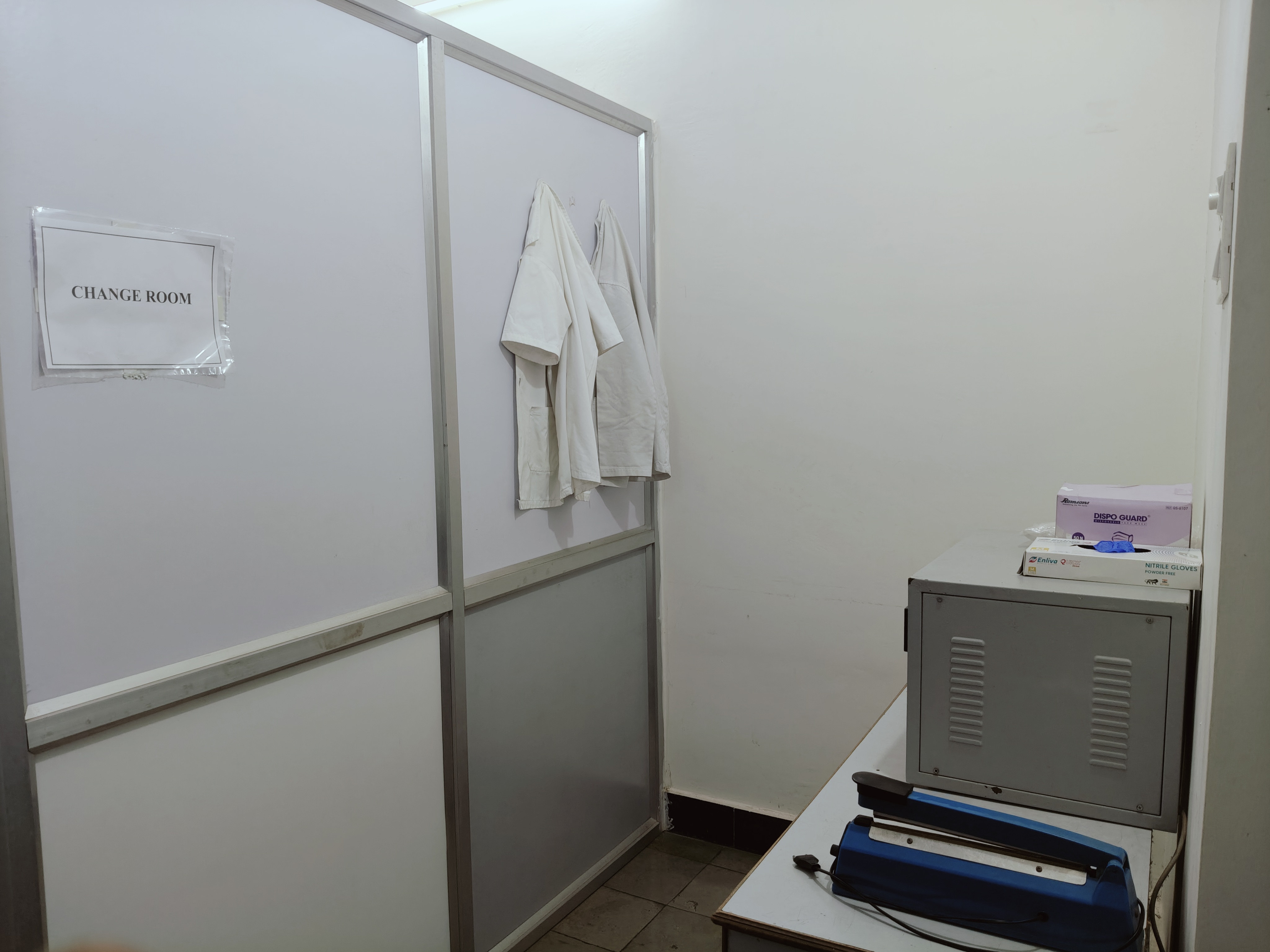
Change Room
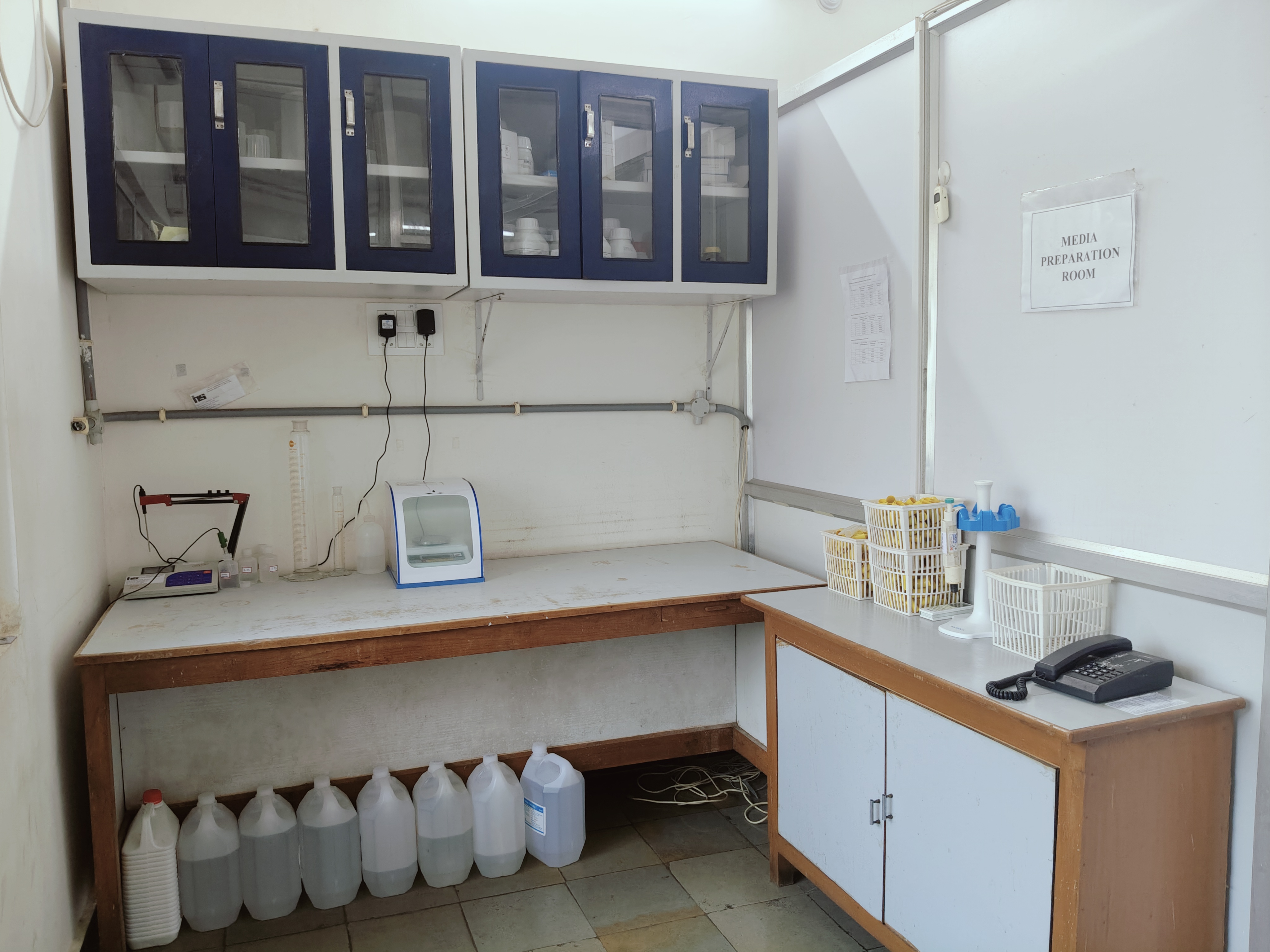
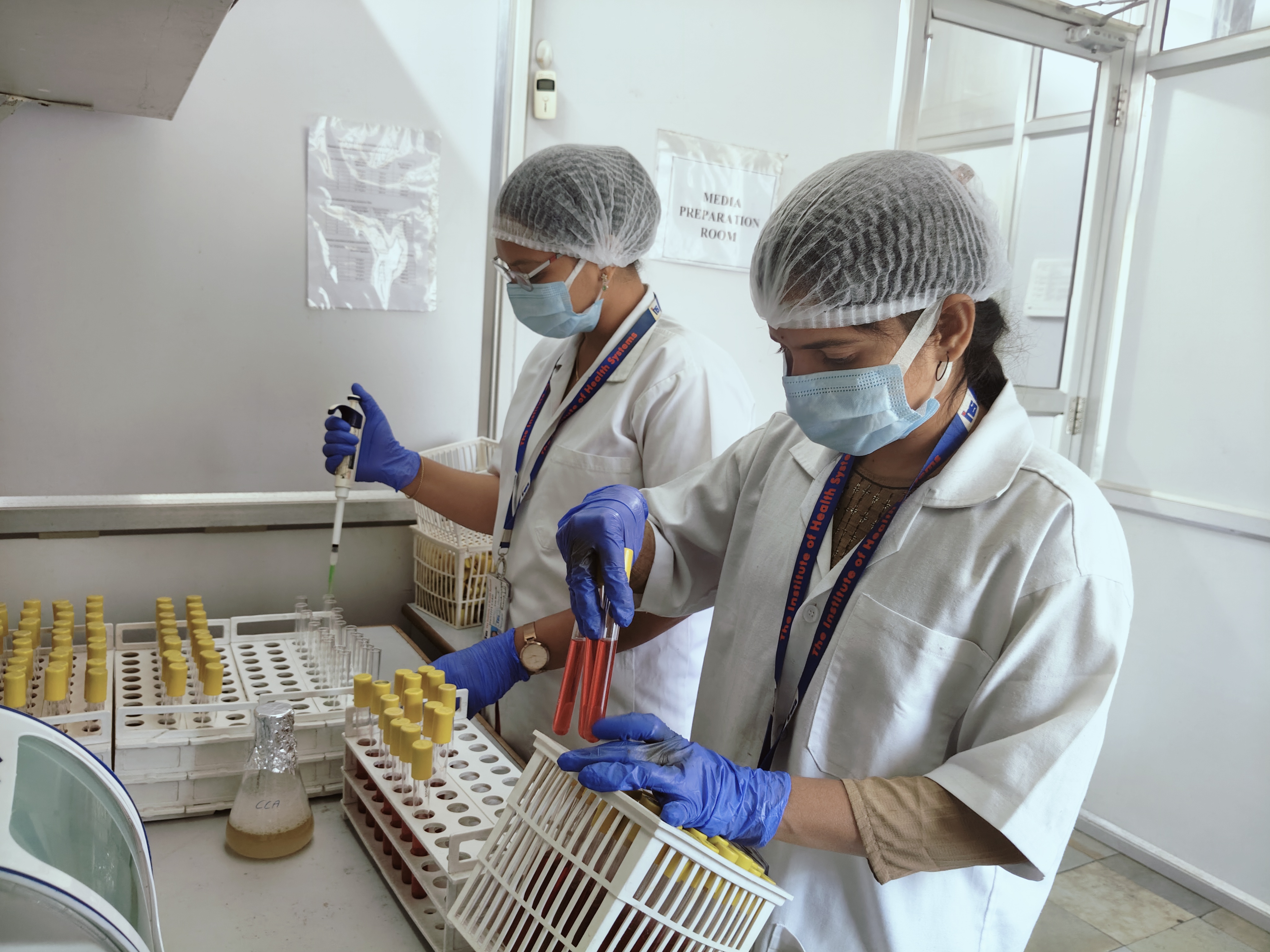
Media Preparation Room
Media Preparation In Progress
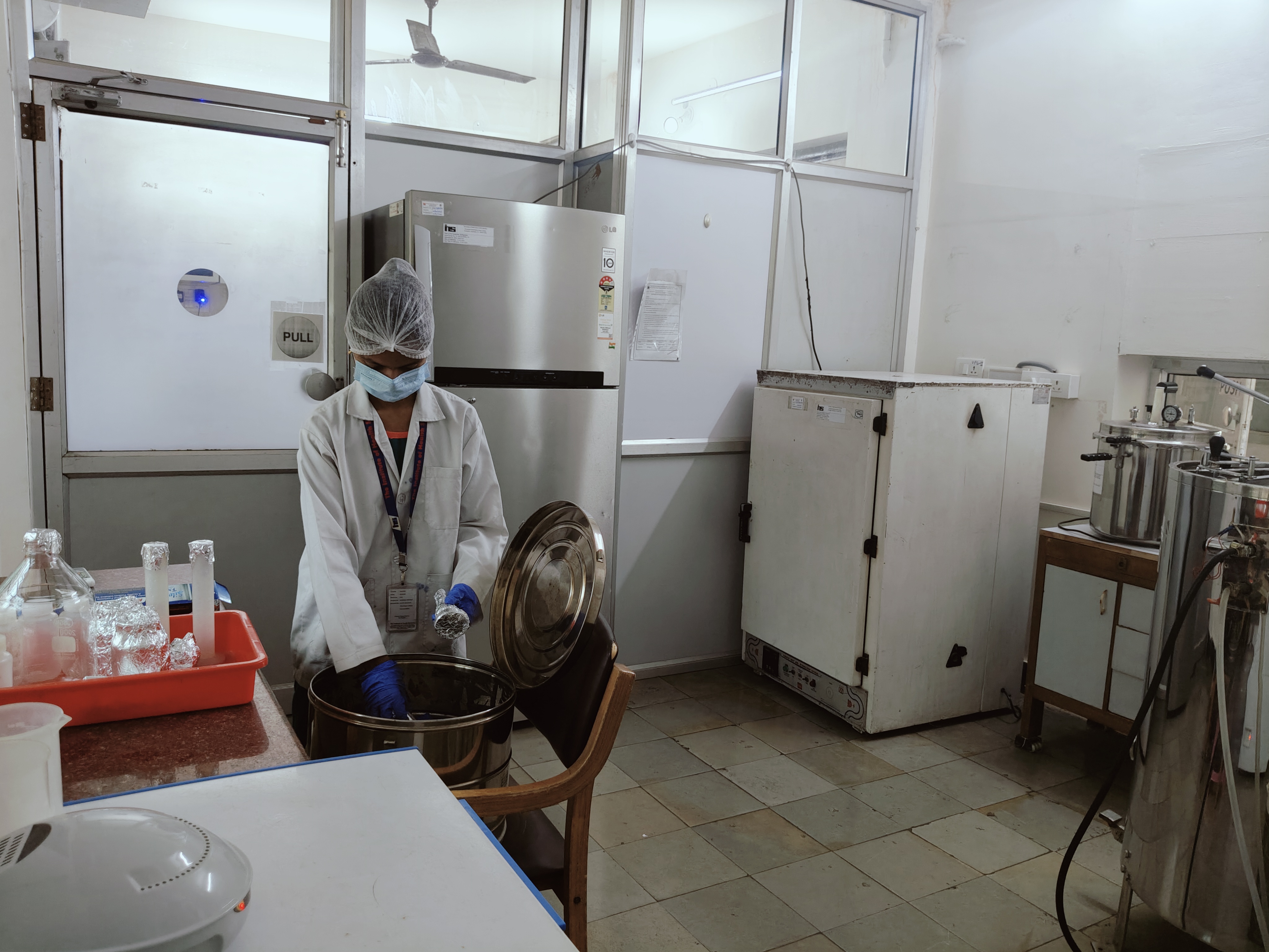
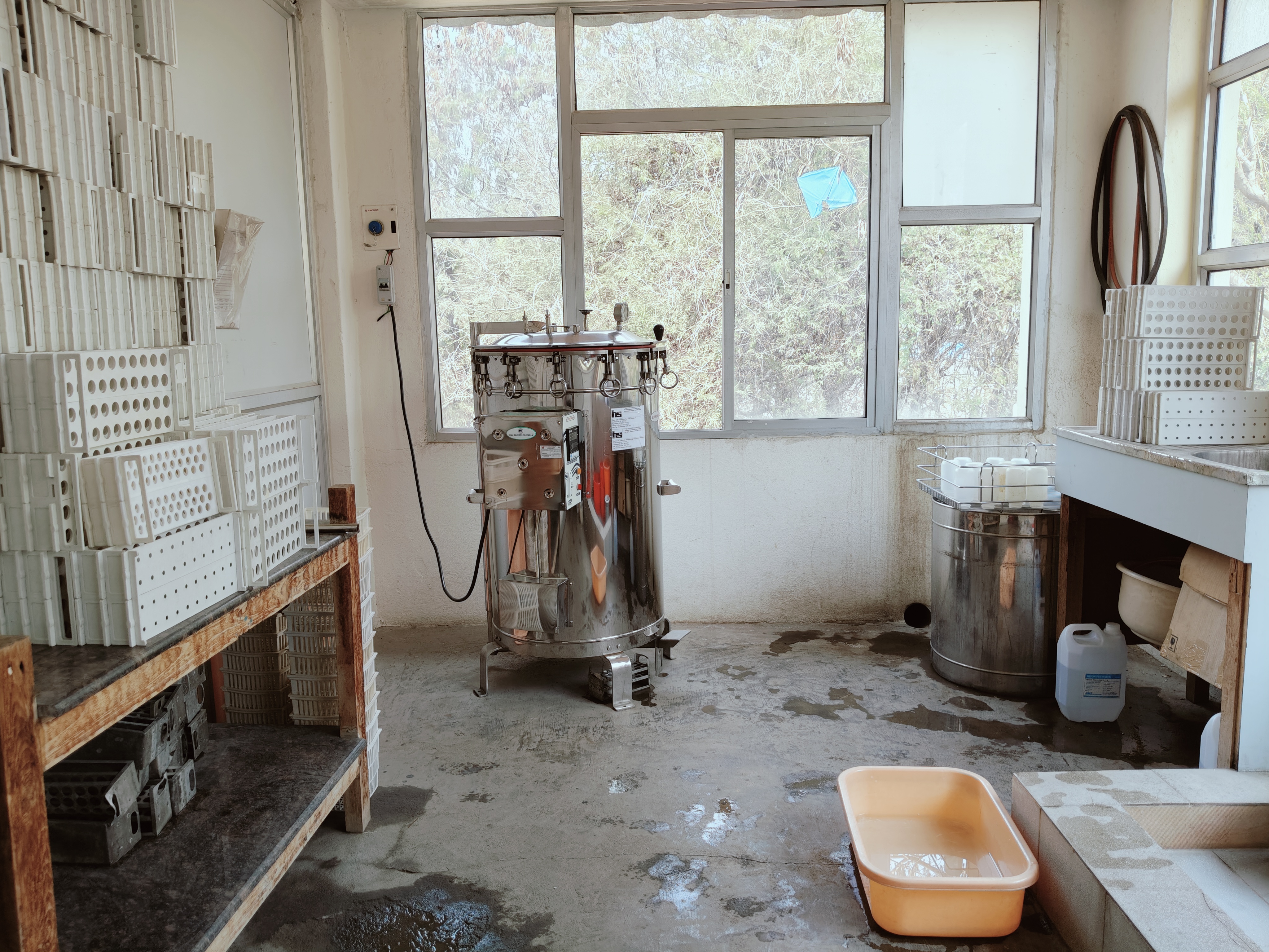
Sterilisation Room
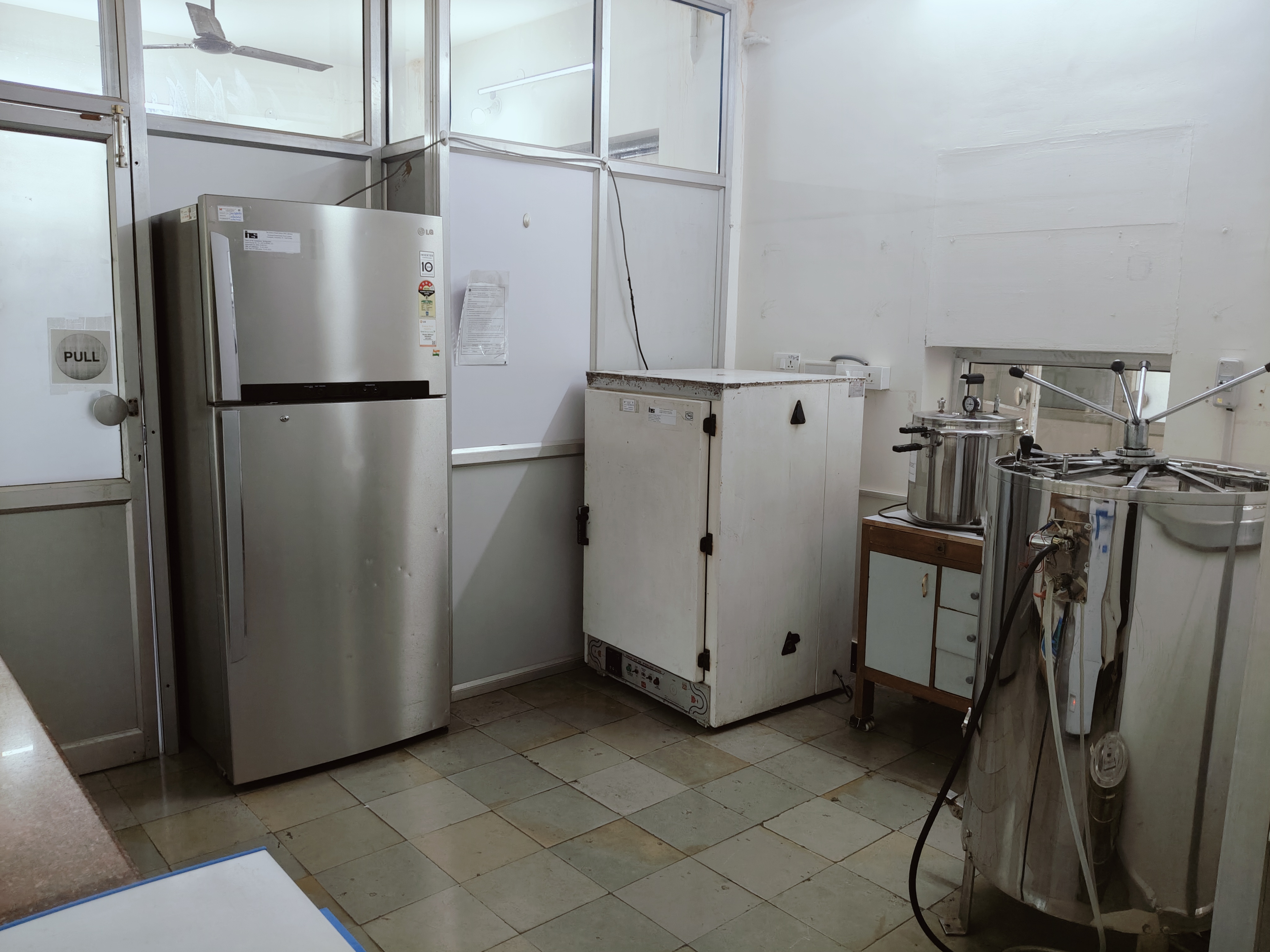
Sterilisation in Progress
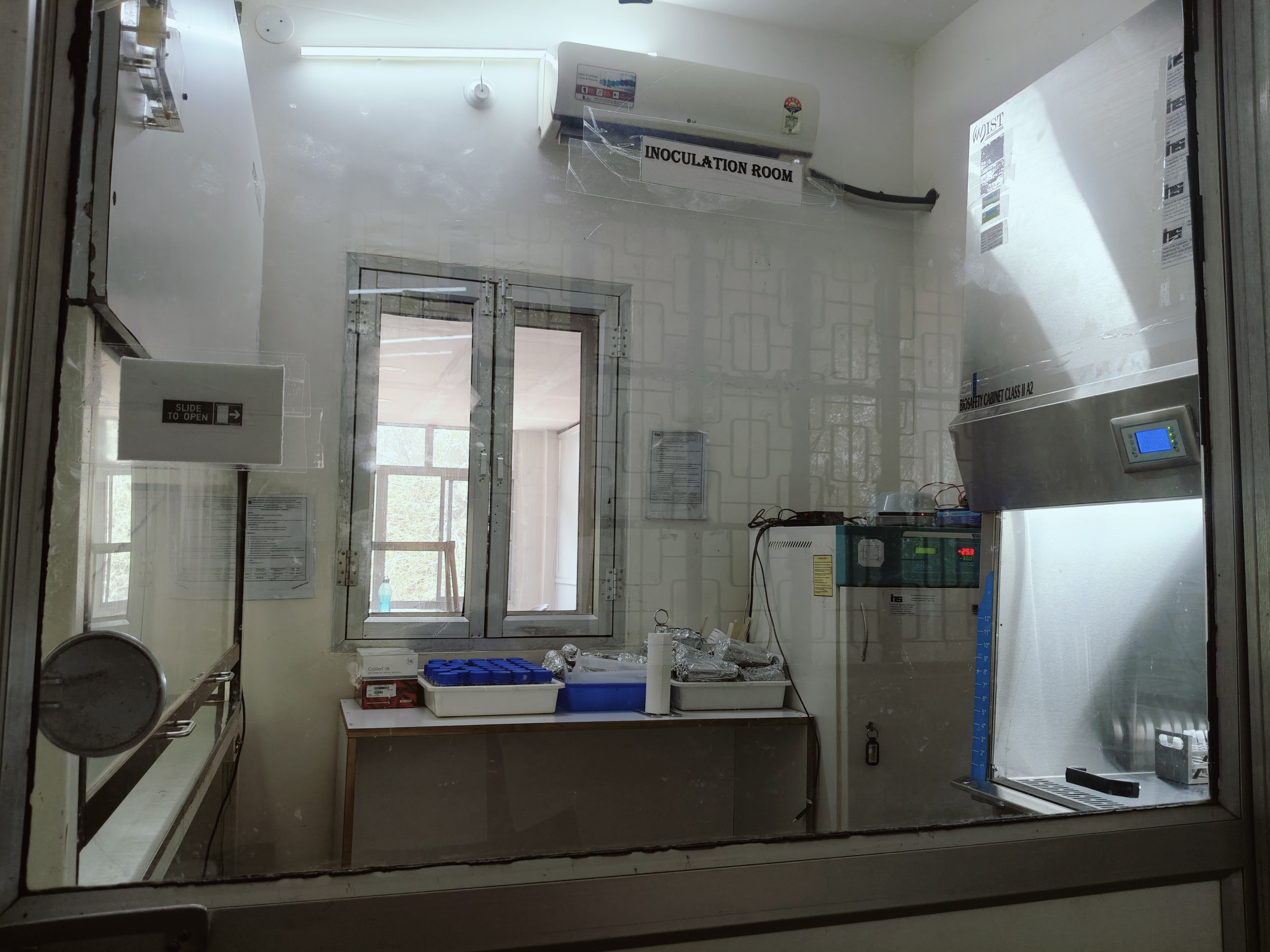
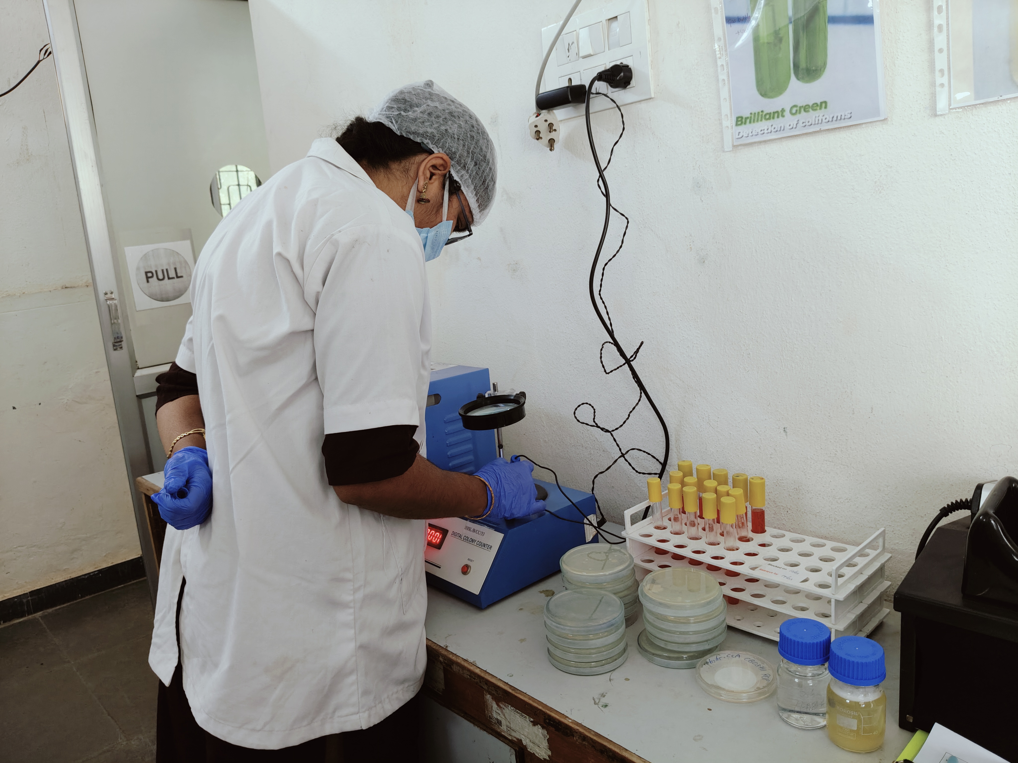
Inoculation Room
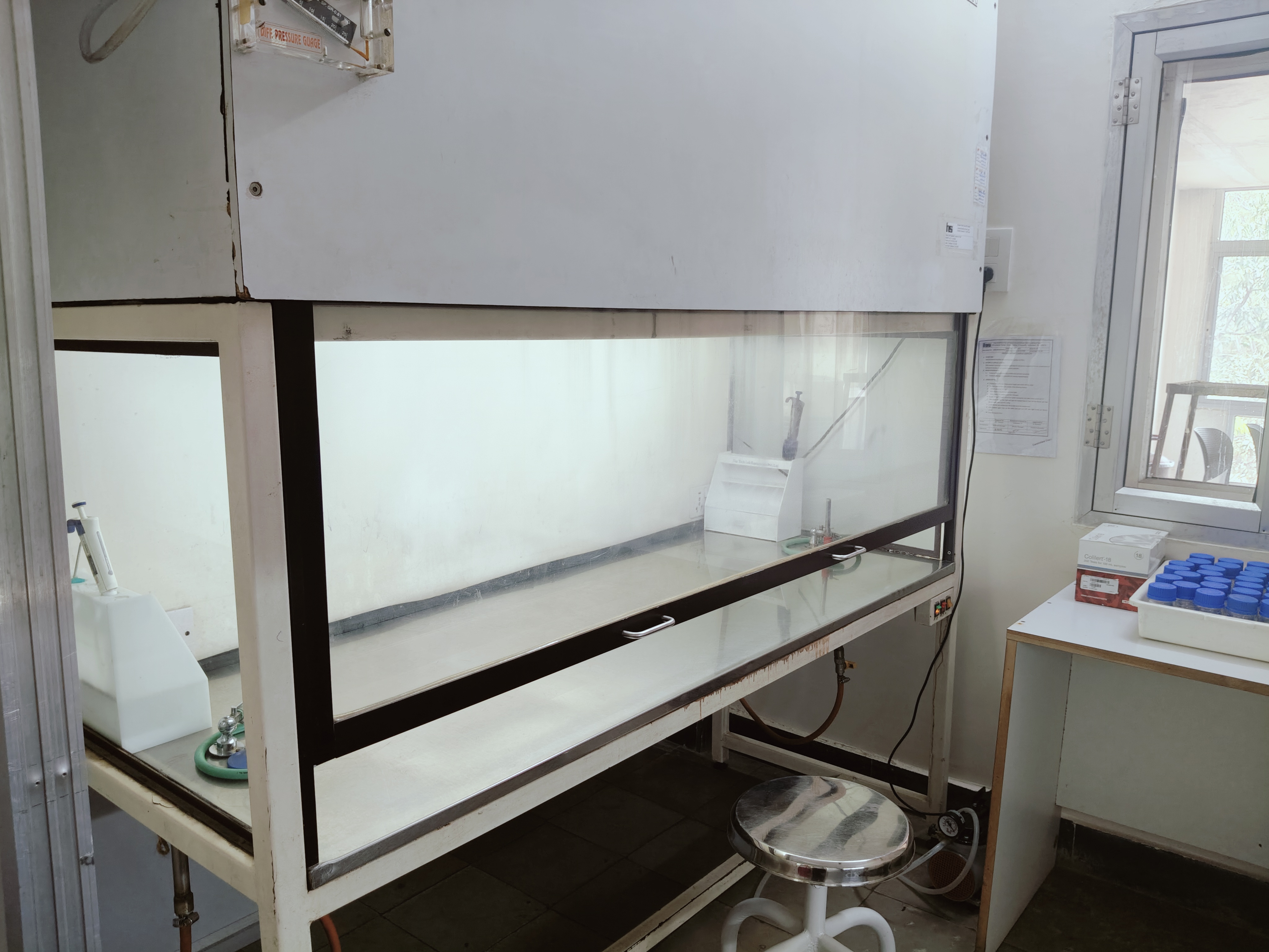
Laminar Airflow Cabinet
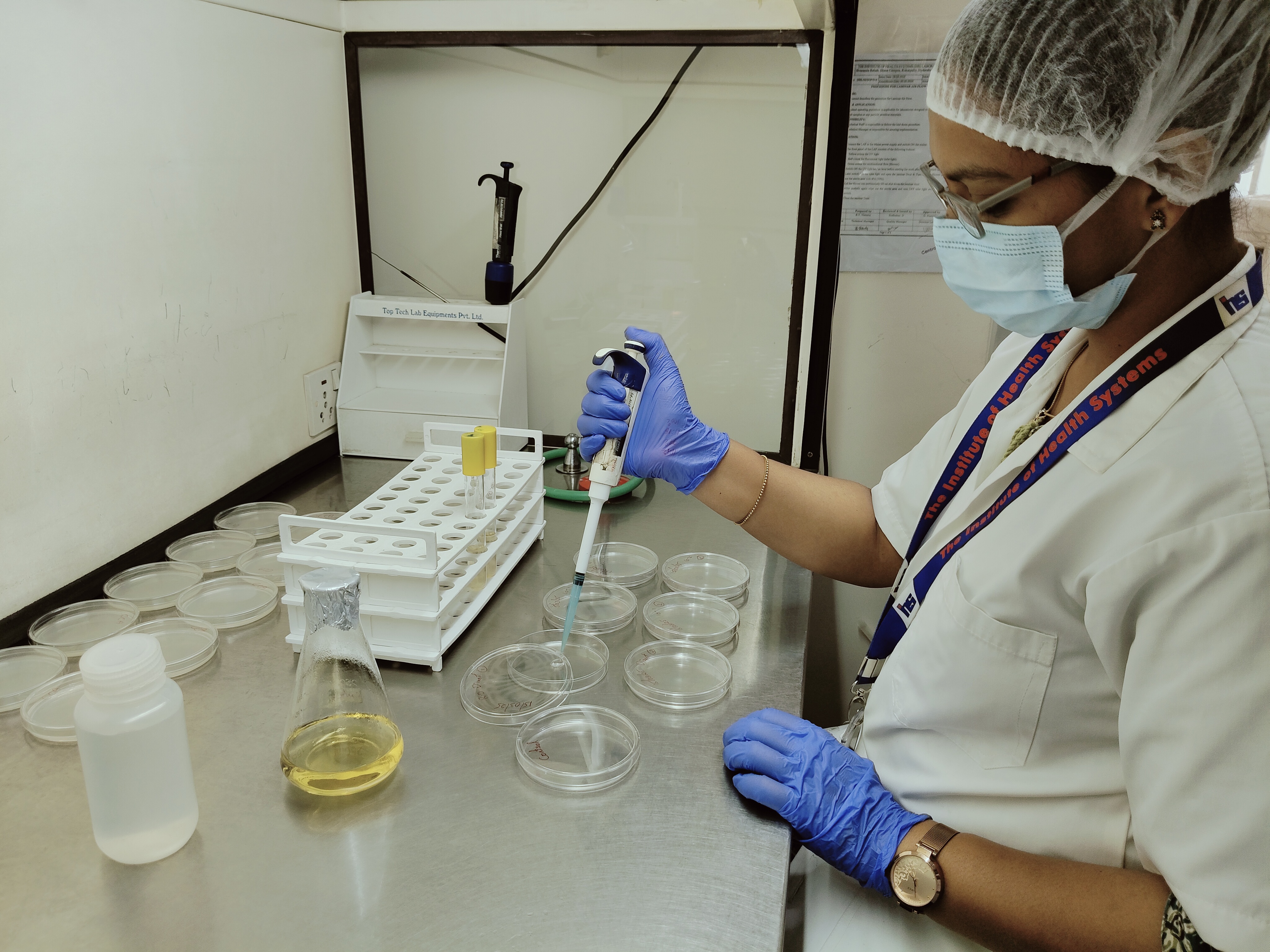
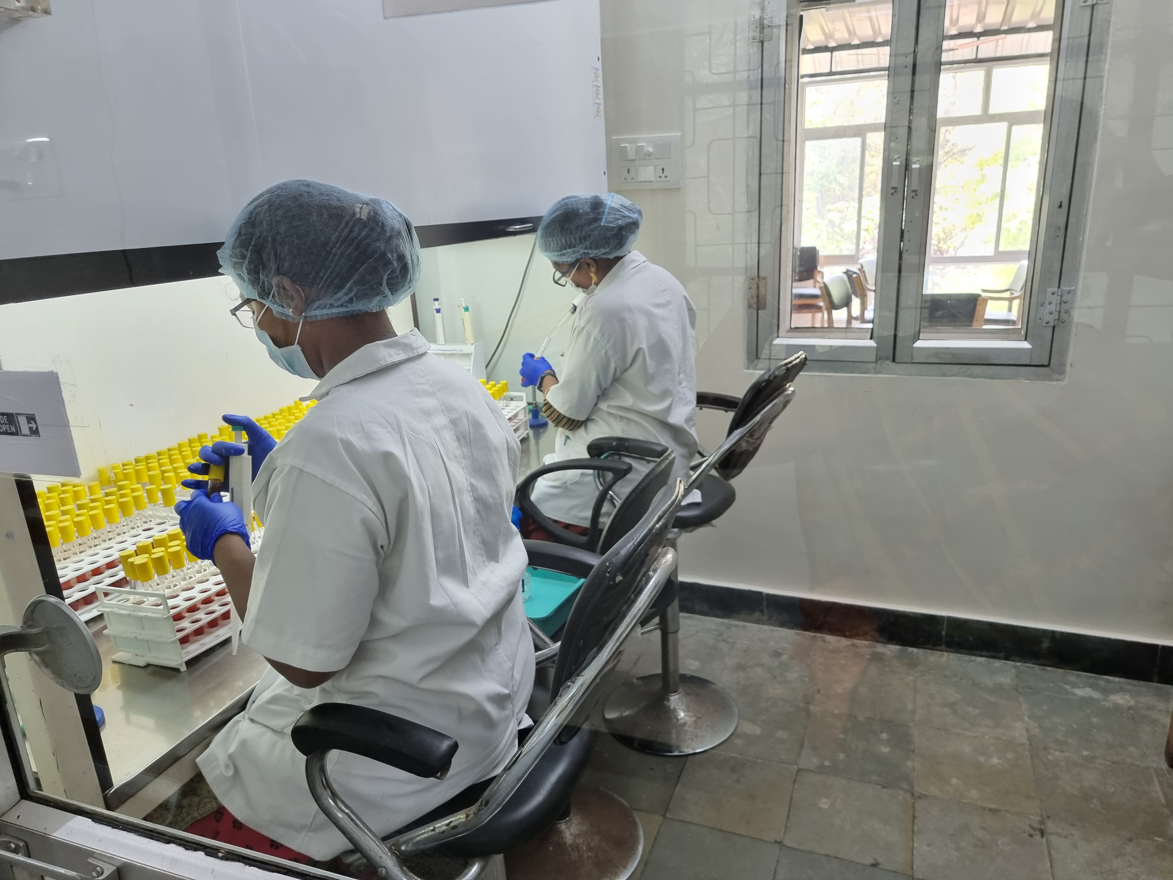
Plating &
Inoculation
in Progress
Multiple Tube -
Inoculation in Progress
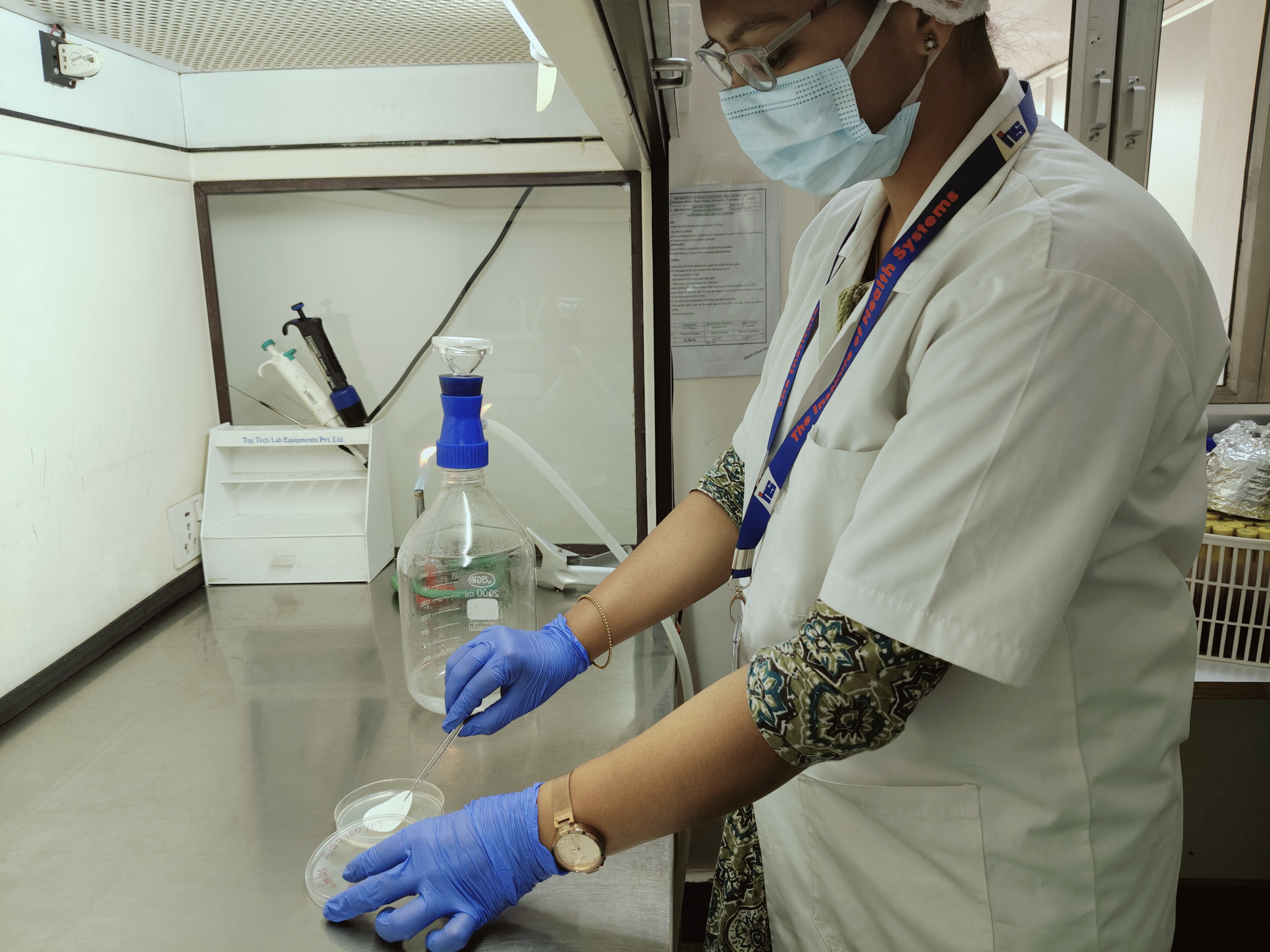
Membrane Filtration
in Progress
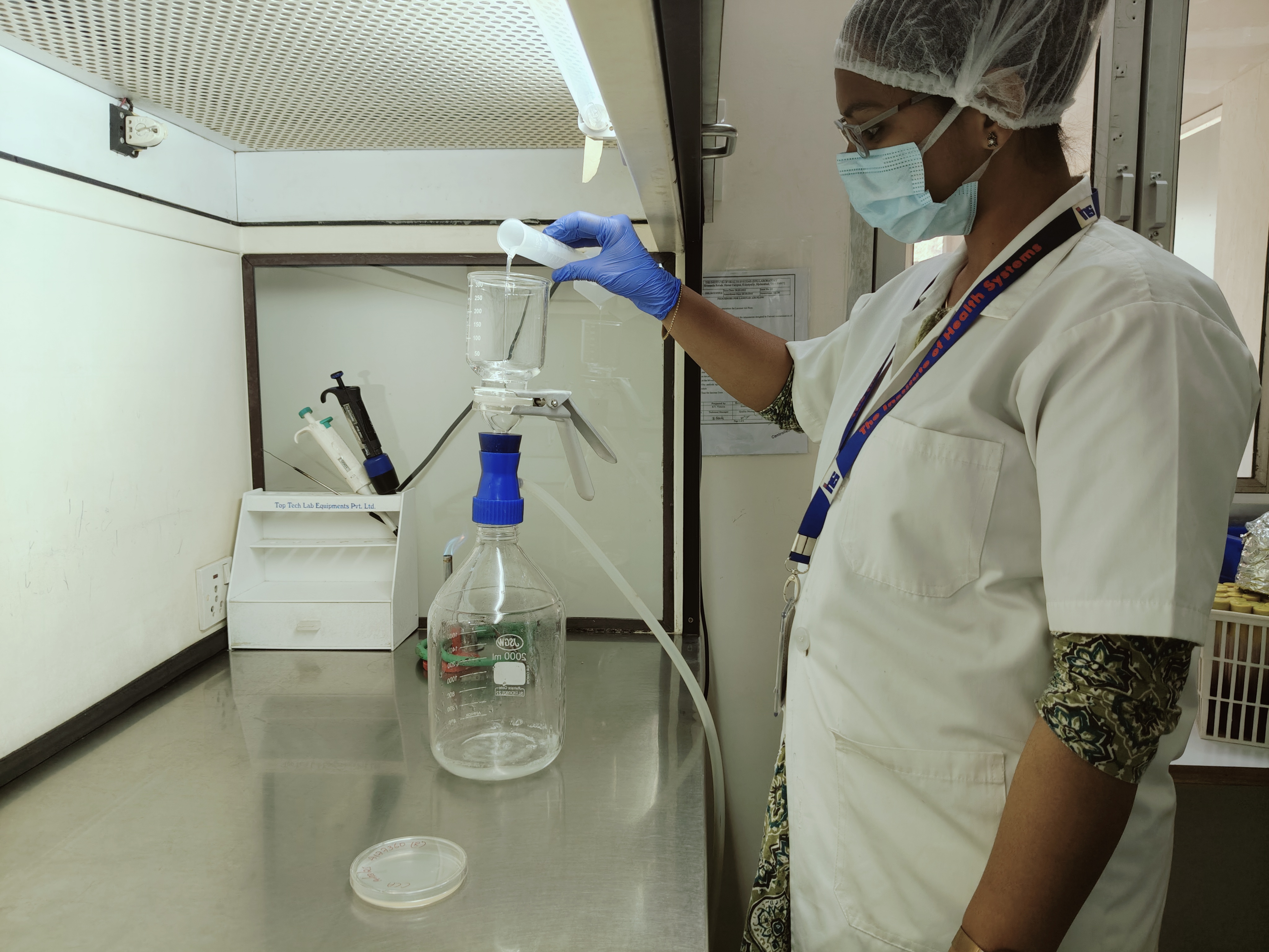
Membrane Filtration &
Inoculation in Progress
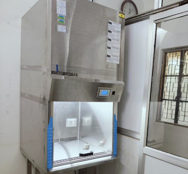
Biosafety
Cabinet
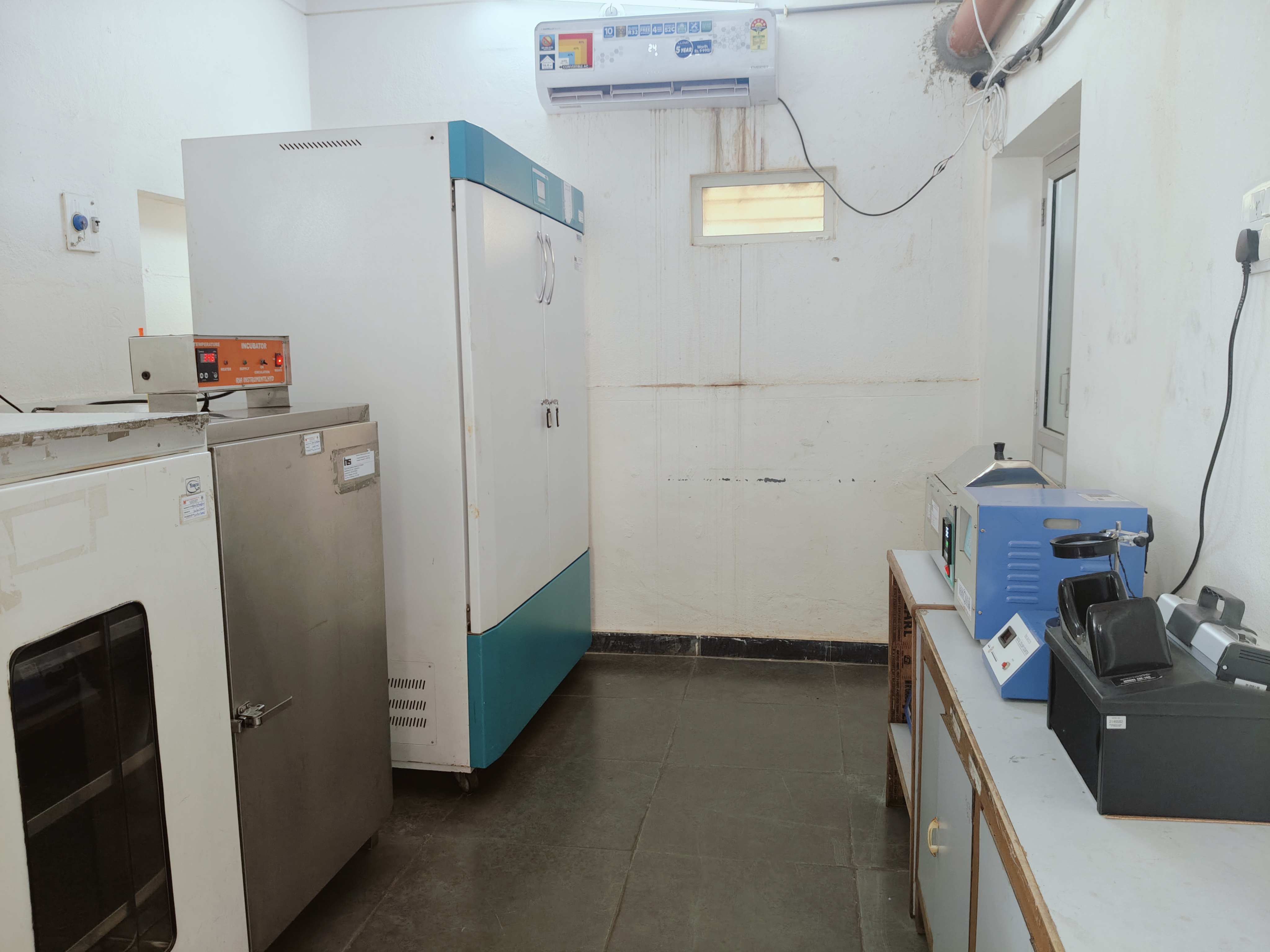
Incubation Room, View-1
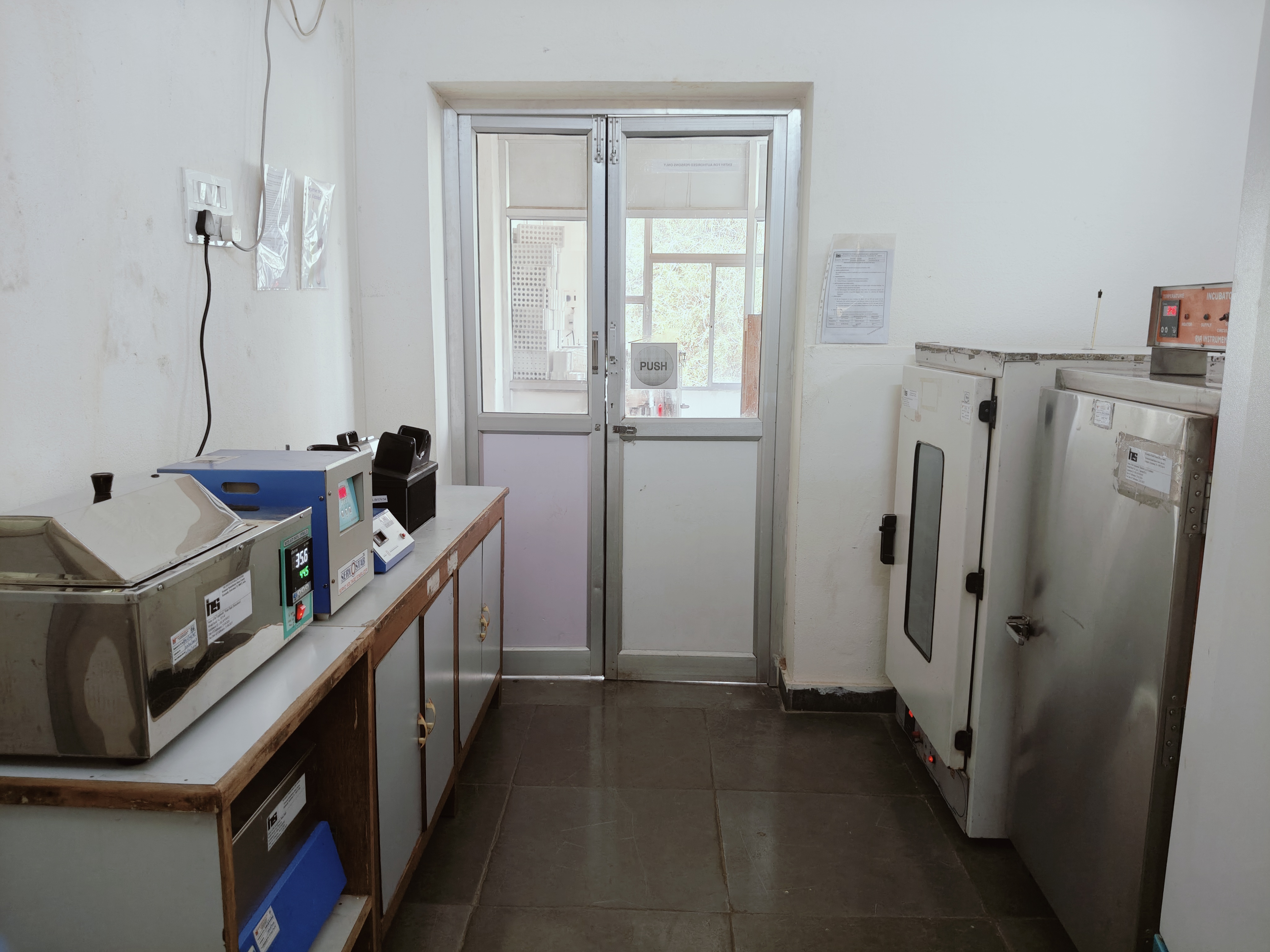
Incubation Room, View-2
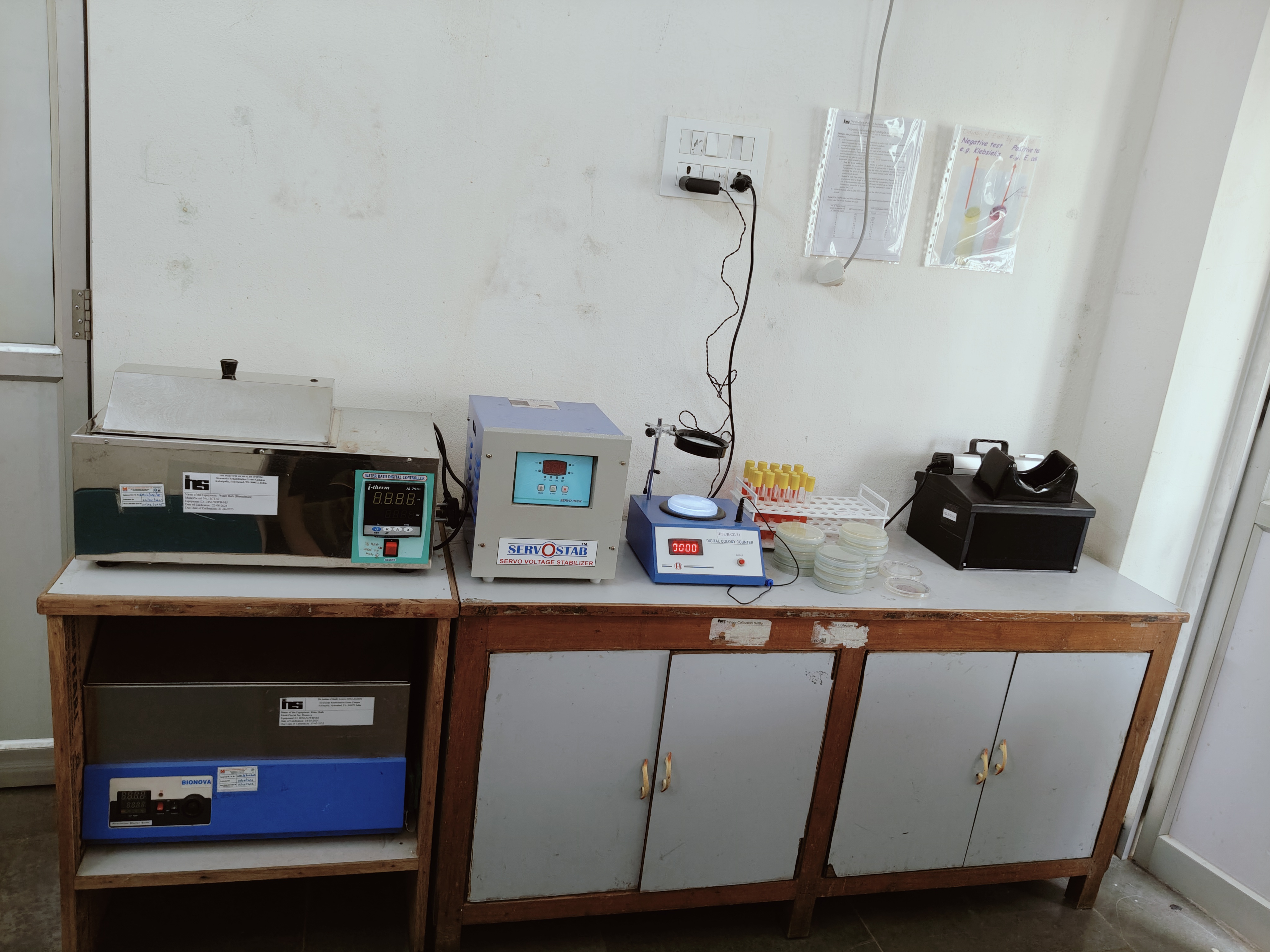
Observation Station
Observation In Progress, View-1
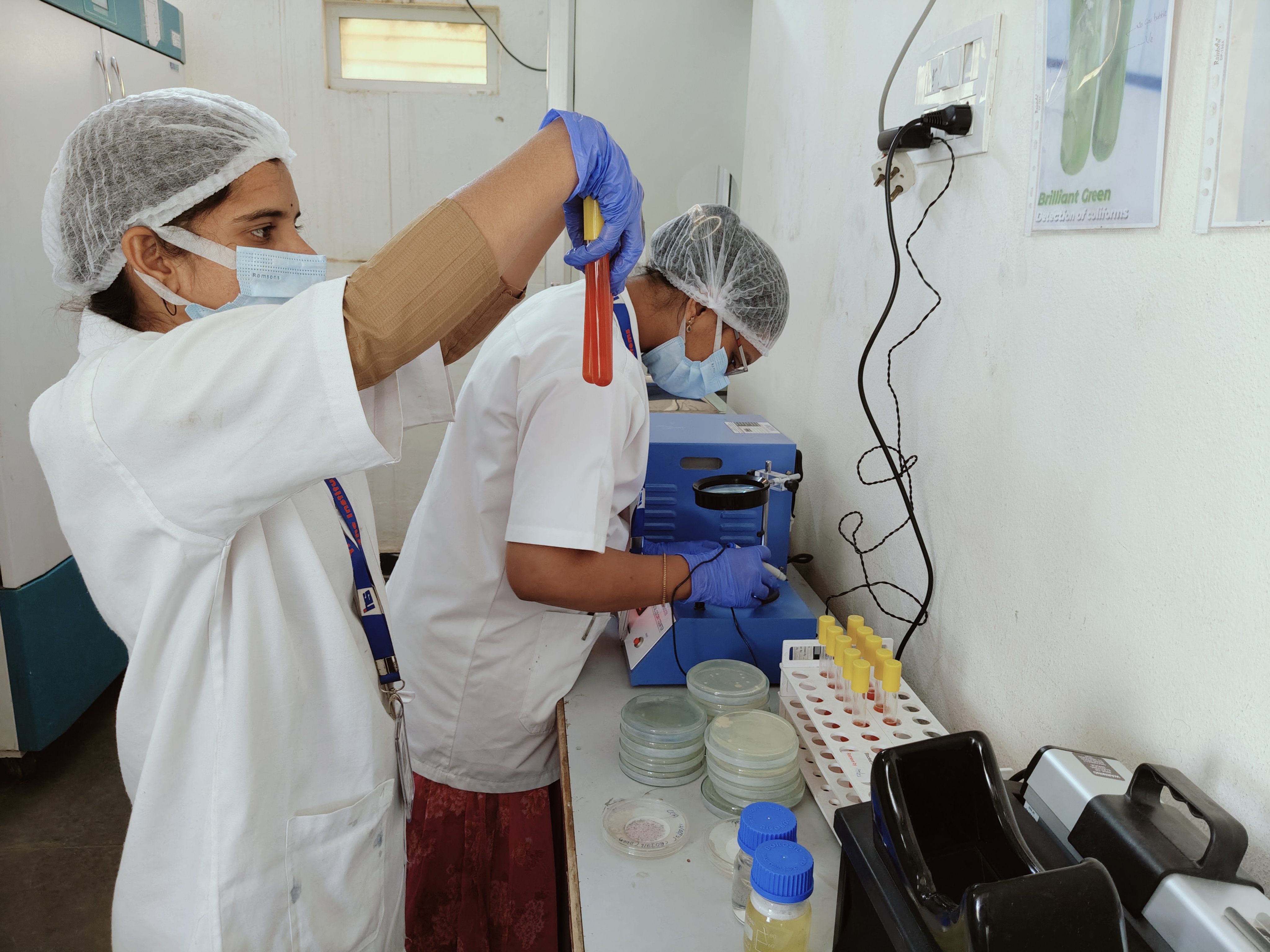
Observation In Progress, View-2
Decontamination Room
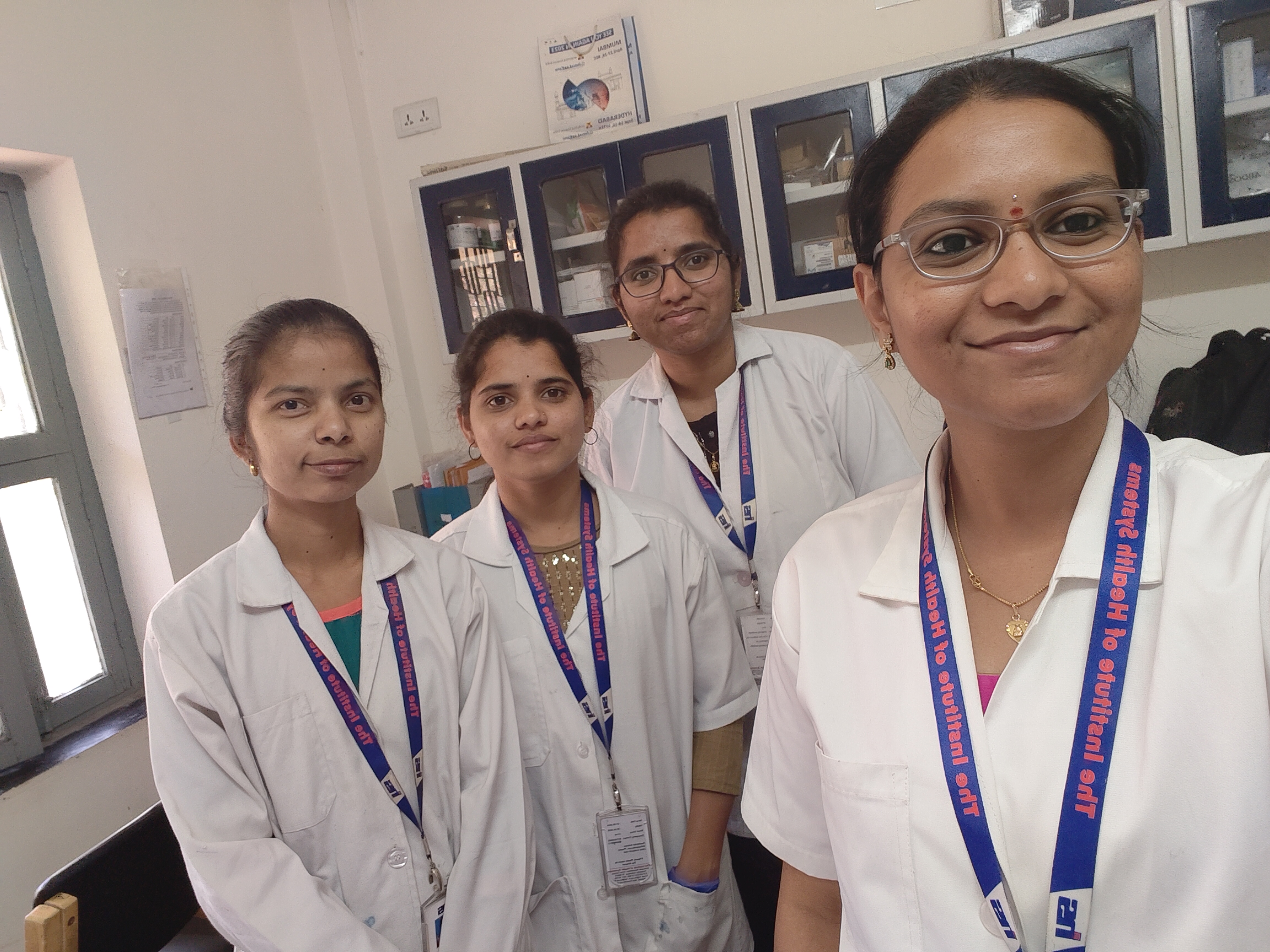
Our Biolab Team
The BioLab is equipped to analyse and test for microbial contamination of water. Various plate count tests are available to estimate bacterial colony forming units (cfu), which is a measure of the total bacterial population in a water sample. Multiple tube dilution technique is used to estimate most probable number (MPN) of total coliforms, some of which would indicate environmental and faecal contamination of water. Confirmatory tests are available to identify indicator organisms associated with environmental and sewage contamination. The bacterial endotoxin assay is available to test the level pyrogens (endotoxins) in water for dialysis.
The plate count methods rely on growth of bacterial colonies on a variety of nutrient mediums incubated at specific temperature for various durations, so that colony becomes visible to the naked eye. But too many colonies, some of which may coalesce with each other would be difficult to count. Hence multiple Petridishes with chosen nutrient medium are inoculated with different dilutions of test sample and the colony count is multiplied by the dilution factor.
In the most probable number (MPN) tests, rows of tubes containing lactose broth are inoculated with water samples measuring 10 ml, 1 ml, and 0.1 ml. During incubation, coliform organisms, if present, would produce gas. A small inverted tube (Durham tube) is placed in each test tube to capture as gas is produced by coliform bacteria. Durham tubes float when filled with gas. The number of test tubes in each row, showing definite signs of gas production is identified and a statistical table is used to arrive at the point estimate and range of coliform bacteria in the sample.
The bacterial endotoxin test (BET), based on the fact that the blood of the horseshoe crab (Limulus or Tachypleus) gels or clots when it comes in contact with endotoxin. The Limulus amebocyte lysate (LAL) reagents are used to test for endotoxin using gel-clot method.

The IHS Laboratory is accredited by the National Board of Accreditation for Testing and Calibration Laboratories (NABL) India.
Lab Id: T-4179; Certificate Number: TC-15240; Issue Date: 02-01-2025; Valid Until: 01-01-2029.
The IHS Laboratory is gradually developing itself as an easily accessible Public Health Laboratory for laboratory tests that are relevant to safeguarding and improvement of people’s health. Keeping with this objective, new test services are introduced to meet felt needs. The IHSL follows sound quality assurance mechanisms and adopts standard test methods for all tests including newly introduced tests. However, immediate accreditation by NABL may not be feasible, for various reasons. Thus, at any point of time, the laboratory would have a set of NABL accredited parameters, and other parameters for which NABL accreditation is not yet available. To avoid any misrepresentation, the IHSL we adopt a strict policy of using NABL symbol in test reports only if all test results included in the report are within the scope the accreditation. For more details see;
The IHSL Policy for Use of NABL Symbol and Claim of NABL Accreditation on Test Reports.
BioLab - Scope of Service
Test Parameters Accredited by NABL, vide certificate number TC-7658, date 02-01-2025, valid until 01-01-2029.
| Sl | SvCd | Parameter | Test Material | Test Method in Brief |
|---|---|---|---|---|
| Plate Counts: | ||||
| Common Procedure: Step-1, Media Preparation: Choose applicable media. Estimate required volume @ 150ml/sample. Suspend required amount of ready-to-use media powder in distilled water & heat the mixture until the agar is completely dissolved. Autoclave the mixture at 121°C for 15 min & let it cool to 45°C. Maintain molten media at 43±2°C for plating. Step-2, Inoculation & Plating: Label required number of sterile Petri plates, including one for blank. Plan & prepare required serial dilutions of the sample, to grow about 30 to 300 colonies on a plate. Mix sample/dilutions well, before inoculation. Ready two plates for each dilution. Pipette 1.0 ml of sample into sterile Petri dish. Pour 15 ml melted media over the sample, tilt & rotate gently to mix sample with molten media. Let the agar-sample mixture solidify at room temperature. Step-3, Incubation: Identify the incubator that is set to appropriate temperature for the particular plate count experiment. Place the plates upside down (inverted), preferably on a separate shelf and incubate for specified duration. Step-4, Counting & Reporting: If no colony is formed in any plate, report "No Growth". Otherwise, proceed with counting. Count colonies formed on surface and within agar, using counting aid (colony counter). Disregard plates with colonies outside the counting range (30 - 300). Report result as the average of all plates falling within counting limit. If number of colonies in undiluted sample < 30 report TFTC. If colonies in all plates are confluent (>300), report TNTC. Reporting Unit: CFU/ml. | ||||
| SPC | Standard Plate Count | Water for Food Processing, Groundwater, Drinking Water, Surface Water, Irrigation Water, Swimming Pool Water. | IS 1622 - 1981, RA 2019, cl 3.2: Step-1: Applicable media: Nutrient Agar. Amount of ready-to-use nutrient agar for step-1: 4.2g in 150ml water for 1 sample (28g/1L); Final pH at 25°C=7.4±0.2". Step-2: Follow common procedure. Step-3: Incubation: At 37°C for 24h. Step-4: Follow common procedure. Reporting Unit: CFU/ml. | |
| HPC | Heterotrophic Plate Count | Water for Food Processing, Drinking Water, Groundwater, Surface Water, Irrigation Water, Swimming Pool Water. | IS 5402 - 2012 RA 2018 / ISO 4833 - 2003: Step-1: Applicable media: Plate count agar (PCA). Amount of ready-to-use PCA for step-1: 3.525g in 150ml water for 1 sample (23.5g/1L); Final pH at 25°C=7.0±0.2". Step-2: Follow common procedure. Step-3: Incubation at 30°C for 72h. Step-4: Follow common procedure. Reporting Unit: CFU/ml. | |
| AMC | Aerobic Microbial Count | Purfied Water, Water from Purifier. | IS 5402 - 2012 RA 2018 / ISO 4833 - 2003 rw IS 14543 - 2024: Step-1: Applicable media: Plate count agar (PCA). Amount of ready-to-use PCA for step-1: 3.525g in 150ml water for 1 sample (23.5g/1L); Final pH at 25°C=7.0±0.2". Step-2: Follow common procedure. Step-3: Incubation at 37±1.0°C for 24h. Step-4: Follow common procedure. Reporting Unit: CFU/ml. | |
| CPC | Cold Plate Count | Purified Water. | IS 5402 - 2012 RA 2018 / ISO 2003 rw IS 14543 - 2024: Step-1: Applicable media: Plate count agar (PCA). Amount of ready-to-use PCA for step-1: 3.525g in 150ml water for 1 sample (23.5g/1L); Final pH at 25°C=7.0±0.2". Step-2: Follow common procedure. Step-3: Incubation at 21±1.0°C for 72h. Step-4: Follow common procedure. Reporting Unit: CFU/ml. | |
| TVC | Total Viable Count | Water for Medicinal Purposes. | IS 17646 Part(3) - 2021 / ISO 23500-3, 2019: Step-1: Applicable media: Tryptic soy agar (TSA). Amount of ready-to-use TSA powder for step-1: 6.75g in 150ml water for 1 sample (45.0g/1L). Step-2: Follow common procedure. Step-3: Incubation at 36±1.0°C for 48h. Step-4: Follow common procedure. Reporting Unit: CFU/ml. | |
| Enzyme Substrate Autoanalysis for Detection of Coliforms & E. coli, Standard Method 9223: | ||||
| Principle: Total coliforms produce the enzyme β-ᴅ-galactosidase which cleaves the chromogenic substrate ONPG and releases yellow chromogen. Most E. coli strains produce the enzyme β-glucoronidase, which cleaves a fluorogenic substrate MUG to release fluorgen. Thus, naked eye observation of yellow colour in culture media containing ONPG indicates presence of total coliforms. Similarly, observation bluish fluoroscence under long-wavelength (365-366nm) ultraviolet (UV) chamber indicates presence of E. coli. Hence, media containing both substrates namely, ONPG and MUG can be used to identify total coliforms and E. coli.
Colilert-18 powder contains; (a) chromogenic substrates ONPG and CPRG, respectively to detect the enzyme β-ᴅ-galctosidase, which is produced by total coliform bacteria; and (b) MUG to detect the enzyme β-glucoronidase, which is produced by E. coli. |
||||
| Common Procedure: Step-1, Prepare Incubation Bottle: Use 100 ml transparent, nonfluoroscent, borosilicate glass bottle, having headspace above 100ml-mark and fitted with screw cap. The have headspace above 100 ml mark is required for proper mixing of media with sample. Sterilise the incubation bottle. Step-2, Inoculation: Pour contents of premeasured & sterile ONPG+MUG substrate powder in Colilert-18 packet into the incubation bottle. Fill sample upto 100ml mark. Do not overfill. Close cap tightly and shake the bottle, vigorously back and forth at least 25 times to properly mix the media with the sample. Step-3, Incubation: Prewarm the bottle in water bath at 35±0.5°C for 20 minutes to bring it to incubation temperature. Place bottle in the incubator within 30 minutes of mixing the medium & sample. Then, Incubate at 35±0.5°C for ≥18h. | ||||
| TCDES | Total Coliform Detection by Enzyme Substrate | Drinking Water, Groundwater, Surface Water. | APHA Standard Methods 24th Ed. 9223 B. Enzyme Substrate Test, 4.a Presence-absence (P/A) procedure: Steps 1-3: Follow common procedure. Step-4, Observation, Reincubation & Reporting: If the sample is yellow or darker yellow than the comparator, it is positive for total coliforms. If unclear after 18 hours, incubate up to 4 more hours; if it turns yellow within this time, it is positive. Colilert-18 can be incubated for up to 22 hours; negative results remain valid after 22 hours, but positive results do not. | |
| ECDES | E. coli Detection by Enzyme Substrate | Drinking Water, Groundwater, Surface Water. | APHA Standard Methods 24th Ed. 9223 B. Enzyme Substrate Test, 4.a Presence-absence (P/A) procedure: Steps 1-3: Follow common procedure. Step-4, Observation & Reporting: Observe in Fluorescence Analysis Cabinet. If the sample is yellow or darker and shows blue fluorescence under an ultraviolet lamp as bright as or brighter than the comparator, it is positive for E. coli. If the fluorescence isn’t clear after 18 hours, incubate up to 4 more hours. If the fluorescence intensifies to match or exceed the comparator within this time, it is positive for E. coli. | |
| Enzyme Substrate based Probable Number of Coliforms & E. coli, Standard Method 9223 B.4.b: | ||||
| Common Procedure: Step-1, Prepare Incubation Bottle: Use 100 ml transparent, nonfluoroscent, borosilicate glass bottle, having headspace above 100ml-mark and fitted with screw cap. The have headspace above 100 ml mark is required for proper mixing of media with sample. Sterilise the incubation bottle. Step-2, Inoculation: Pour contents of premeasured & sterile Colilert-18 packet into the incubation bottle. Fill sample upto 100ml mark. Do not overfill. Close cap tightly and shake the bottle, vigorously back and forth at least 25 times to properly mix the media with the sample. Step-3, Distribution: Pipette out contents of the bottle into 10 sterile test tubes @ 10 ml in each tube. Step-4, Prewarming: Prewarm the inoculated test tubes in water bath at 35±0.5°C for 20 minutes to bring it to incubation temperature. Place the 10-tube rack inside incubator within 30 minutes of mixing the medium & sample. Then, Incubate at 35±0.5°C for ≥18h. | ||||
| EPNTC | Enzyme Substrate based Probable Number of Total Coliforms | Drinking Water, Groundwater, Surface Water. | APHA Standard Methods 24th Ed. 9223 B. Enzyme Substrate Test, 4.b Multiple-tube procedure: Steps 1-4: Follow common procedure. Step-5, Observation: Observe each tube. If the sample is yellow or darker yellow than the comparator, it is positive for total coliforms. If unclear after 18 hours, incubate up to 4 more hours; if it turns yellow within this time, it is positive. Colilert-18 can be incubated for up to 22 hours; negative results remain valid after 22 hours, but positive results do not. Step-6, Computation of Results: Refer APHA Standard Methods 24th Ed. Table 9221:3,Page 1137. MPN Index and 95% Confidence Limits for all combinations of positive and negative results when Ten 10-mL Portions are used. | |
| EPNEC | Enzyme Substrate based Probable Number of E. coli | Drinking Water, Groundwater, Surface Water. | APHA Standard Methods 24th Ed. 9223 B. Enzyme Substrate Test, 4.b Multiple-tube procedure: Steps 1-4: Follow common procedure. Step-5, Observation: Observe each tube in Fluorescence Analysis Cabinet. If the sample is yellow or darker and shows blue fluorescence under an ultraviolet lamp as bright as or brighter than the comparator, it is positive for E. coli. If the fluorescence isn’t clear after 18 hours, incubate up to 4 more hours. If the fluorescence intensifies to match or exceed the comparator within this time, it is positive for E. coli. Step-6, Computation of Results: Refer APHA Standard Methods 24th Ed. Table 9221:3,Page 1137. MPN Index and 95% Confidence Limits for all combinations of positive and negative results when Ten 10-mL Portions are used. | |
| Multipe Tube Fermentation Tests: | ||||
| MPNTC | MPN (Total coliforms) by multi-tube dilution | Water for Food Processing, Groundwater, Surface Water, Irrigation Water, Swimming Pool Water, Wastewater. | IS 1622 - 1981, RA2019, cl 3.3.1 Multi-tube dilution test (MDT):
Step-1, Media Preparation: Prepare double & single strength MacConkey broth using certified ready-to-use media. Distribute 10 ml of double strength media into 150 x 18 mm tubes; and 10 ml of single strength media into 150 x 15 mm test tubes. Place an inverted Durham's tube into each media tube, plug the test tubes with non-absorbent cotton and autoclave at 115°C for 10 minutes. Step-2, Innoculation: Arrange 5 presterilised double strength MacConkey broth tubes in 1st row; single strength tubes in rows 2 & 3. Innoculate each of the 1st row tubes with 10 ml; 2nd row tubes with 1.0 ml; and 3rd row tubes with 0.1 ml of water sample. Step-3, Incubation: Incubate all tubes at 37°C for 24±2h. Step-4, Observation: Observe & media colour change, if any; position of Durham's tube (bottom/floating), and gas in Durham's tube, if any. Record inference about number of presumptive positive tubes in each row. Step-5, Computation of presumptive total coliforms: Refer Appendix B Table-3 MPN per 100 ml sample and confidence limits using 5 tubes of 10 ml, 5 tubes of 1 ml and 5 tubes of 0.1ml. |
|
| FCDMT | Fecal (Thermotolerant) coliforms Detection by multi-tube dilution. | Water for Food Processing, Groundwater, Surface Water, Irrigation Water, Swimming Pool Water, Wastewater. | IS 1622 - 1981, RA2019, cl 3.3.1.2b(i) & 3.3.3: Continue after observation of results for MPN (Total coliforms). Subculture, by transferring one or two loopful from lowest dilution presumptive positive tubes to a tube of BGLB broth and incubate in water bath at 44.5°C for 24±2h. Any amount of gas formation in the Durham's tube indicates fecal coliforms. If negative, reincubate and examine again at 48±2h. | |
| ECDMT | E. coli Detection by multi-tube dilution. | Drinking Water, Water for Food Processing, Water for Dialysis, Purified Water, Groundwater, Irrigation Water, Surface Water, Swimming Pool Water, Wastewater. | IS 1622 - 1981, RA2019, cl 3.3.4: Continue after observation of results for Fecal coliforms FCDMT. Subculture, from Fecal coliform positive BGLB tube, in tryptone water at 44.5°C for 24h. Add a few drops of Kovac's reagent. Pink colour indicates E. coli positive & yellow means negative. | |
| MPNFC | MPN (Fecal Coliforms) by multi-tube dilution. | Water for Food Processing, Groundwater, Surface Water, Irrigation Water, Swimming Pool Water, Wastewater. | IS 1622 - 1981, RA2019, cl 3.3.3.2: Continue after observation of results for MPN (Total coliforms). Subculture, from all presumptive positive tubes of coliform test into as many BGLB tubes at 44.5℃ for 24±2h in water bath. Observe for gas formation (FC +ve) or lack of it (FC -ve). Refer Appendix B Table-3 to estimate MPN(Fecal coliforms) per 100 ml sample. | |
| MPNEC | MPN (E. coli) by multi-tube dilution. | Water for Food Processing, Groundwater, Surface Water, Irrigation Water, Swimming Pool Water, Wastewater. | IS 1622 - 1981, RA 2019, cl 3.3.4: Continue after observation of results for MPN (Fecal coliforms). Subculture, from all positive tubes of BGLB broth, in as many tubes of tryptone water at 44.5℃ for 24±2h in water bath. Add few drops of Kovac's reagent into each tube to test for indole production (pink) or lack of it (yellow). Refer Appendix B Table-3 to estimate MPN (E. coli) per 100 ml sample. | |
| Membrane Filtration & Chromogenic-agar plating based Detections (MFC): | ||||
| Common Procedure: Step-1, Filtration: Make sure that membrane filtration assembly glassware, clamp(s), forceps & gasket have been sterilised and the vacum pump is connected. Filter required volume (100 / 250 ml) of the sample through a sterile 47mm dia, 0.45µm poresize cellulose membrane. | ||||
| ECDMF | Total coliforms & E. coli Detection by membrane filtration. | Purified Water. | IS 15185, 2016 RA 2021 / ISO 9308-1, 2014: Step-2: Place the membrane on chromogenic coliform agar plate, invert the Petri dish, and incubate at 36 ± 2°C for 21 ± 3 hours. Pink-red colonies indicate total coliforms and Dark blue-violet colonies indicate E. coli. Step-3, Oxidase Test by Filter Paper Spot Method: Using an inert transfer loop pick a well-isolated colony and rub onto a small piece of filter paper. Place 1 or 2 drops of Gordon Mcleod oxidase reagent on the organism smear. Start a stop watch and observe for 30 seconds. If dark blue colour appears within 30 seconds, then oxidase positive. Else, if colour does not change or it takes longer than 30 seconds, then oxidase negative. Total coliforms and E. coli are oxidase negative. | |
| YMDMF | Yeast & Mould detection by membrane filtration. | Purified Water. | IS 5403, 1999 RA 2018 / ISO 7954: Step-2: Place the membrane on a yeast glucose chloramphenicol (YGC) agar plate, invert the Petri dish, and incubate at 25 ± 1°C. Examine colonies on each plate after 3, 4 & 5 days and record the nature of colonies (yeast/mould) detected on each day. Terminate the experiment, after both yeast and moulds are detected, else continue till 5th day and report results accordingly. | |
| SLDMF | Salmonella detection by membrane filtration. | Purified Water. | IS 15187, 2016 RA 2021 / ISO 19250, 2010: Step-2: Place the membrane in 50 mL of buffered peptone water, and incubate at 36 ± 2°C for 18 ± 2 hours. Then, enrich the culture in Rappaport-Vassiliadis medium (0.1 mL culture to 10 mL of RVS broth) at 41.5 ± 1°C for 48 ± 4 hours. Confirm by subculturing on Xylose Lysine Deoxycholate Agar or Brilliant Green/Phenol Red Lactose Agar at 36 ± 2°C for 24 ± 3 hours. Salmonella colonies on XLD agar usually appear red with a black center, while on Brilliant Green/Phenol Red Lactose Agar, colonies are pinkish-white or red with a red halo. Pick typical single colonies from each plate, streak them onto non-selective agar (e.g., Nutrient Agar), and incubate at 36 ± 2°C for 24 ± 3 hours. Finally, perform biochemical confirmation using single colonies from the Nutrient Agar plates on TSI Agar (Salmonella shows alkaline red slants, gas formation, acid yellow butts, and hydrogen sulfide formation with blackening), Urea Agar (Salmonella negative for urea hydrolysis), and L-Lysine Decarboxylation Medium (Salmonella shows a positive purple color). | |
| PADMF | P. aeruginosa detection by membrane filtration. | Purified Water. | IS 13428, 2024 Clause 6.1.5, Annex D: Step-2: Place the membrane in asparagine proline broth medium, and incubate at 37 ± 1°C for 48 hours. Examine for growth and fluorescence under an ultraviolet lamp. For confirmation, subculture from containers showing either fluorescence or growth onto milk agar with cetrimide plate and incubate at 42 ± 0.5°C for 24 hours. Examine the plates for growth, blue-green pigment production, and casein hydrolysis (clearing of the milk medium around the colonies). Colonies showing all these reactions are positive for Pseudomonas aeruginosa. | |
| SADMF | S. aureus detection by membrane filtration. | Purified Water. | IS 5887 (Part 2), 1976 RA 2022: Place the membrane in 50 mL of cooked meat salt medium, and incubate at 37°C overnight (18h). Then, subculture on Baird-Parker medium and incubate at 37°C for 30 hours. S. aureus forms shiny black colonies with or without grey-white margins on Baird-Parker agar. For confirmation, perform a coagulase test by selecting a typical colony and inoculating it into 1 mL of citrated rabbit plasma diluted 1 in 5 with 0.85% saline in a narrow tube. Incubate at 37°C and observe every hour (1-4 hours) for plasma clotting. S. aureus is coagulase positive, showing clotting of the plasma. | |
| Notes: | ||||
Verified & Validated Test Methods Outside the Scope of NABL, Accreditation:
| Sl | SvCd | Parameter | Test Material | Test Method |
|---|---|---|---|---|
| BET | Bacterial Endotoxin Assay (LAL) | Water for Medicinal Purposes, Purified Water. | International Pharmacopoea (Ph.Int.) 12th Ed., 2025, Methods of Analysis, 3. Biological Methods, 3.4 Test for bacterial endotoxins, Method A. The gel-clot technique - Limit test. | |
| FSDMF | Faecal Streptococci detection by membrane filtration | Water for Medicinal Purposes, Purified Water, Packaged drinking water, Drinking water, Swimming pool water, Surface water. | IS 15186, 2002 / ISO 7899-2, 2000: Step-1, Filtration: Common Procedure. Step 2: Place the membrane on Slanetz and Bartley medium and incubate at 36 ± 1°C for 44 ± 4 hours. Presumptive enterococci colonies on Slanetz and Bartley medium appear red, maroon, or pink in the center or throughout the colony. Step 3: For confirmation, transfer the membrane using sterile forceps (without inverting it) onto a plate of Bile-Aesculin-Azide agar, which has been preheated to 44°C. Incubate at 44 ± 0.5°C for 2 hours, then observe the plates immediately.Colonies showing a tan to black color in the surrounding medium indicate a positive reaction and should be counted as Intestinal enterococci/ Faecal streptococci. | |
| SGDMF | Shigella detection by membrane filtration | Water for Medicinal Purposes, Purified Water, Packaged drinking water, Drinking water, Swimming pool water, Surface water. | IS 5887(Part 7), 1999 RA 2005: Step-1, Filtration: Common Procedure. Step 2: Place the membrane in 50 mL of nutrient broth and incubate at 37°C for 18-24h. Then, enrich the culture in Kauffmann-Muller’s Tetrathionate Broth (1 mL culture to 100 mL of KMT broth) at 37°C for 24h. Step 3: Confirm by subculturing on Deoxycholate citrate agar at 37°C for 48h. Shigella colonies on DCA agar appear opaque with a ground-glass appearance and with even margins. Step 4: Pick typical single colonies from each plate, streak them onto MacConkey agar plate and incubate overnight at 37°C. Step 5: Transfer pure colonies onto non-selective agar (e.g., Nutrient Agar), and incubate at 36 ± 2°C for 24 ± 3 hours. Step 6: Perform biochemical confirmation using single colonies from the Nutrient Agar plates on TSI Agar (Shigella shows negative TSI for H2S), Urea Agar (Shigella shows negative for urea hydrolysis) and Oxidase test (Shigella is oxidase negative). | |
| Notes: The laboratory has verified and developed the test method in accordance with concerned standard test method, and validated the same through professional testing but is yet to apply for expansion of NABL accreditation. | ||||
Water Sample Collection Bottles for Bacteriological Analysis:
| SBCd | Bottle Name | Details | Volume |
|---|---|---|---|
| CSBF | Clean Sterile Bottle for Freshwater | Clean sterile rigid polypropylene bottle without any additive for Freshwater / purified water known to be free of chlorine. | 250ml |
| SBTD | Sterile Bottle with Thiosulfate for Drinking / Fresh water. | First clean, rinse and dry 250 ml rigid pollypropylene bottles. Then, add 10 drops (0.5ml) of 1.25% w/v anhydrous Na2S2O3 solution to neutralise up to 5ppm residual chlorine in 250 ml of drinking / fresh water for bacteriological analysis. Sterilize in audoclave. Fix label and pack in sealed polythene bags. [APHA Standard Methods 9060.] | 250ml |
| SBTP | Sterile Bottle with Thiosulfate for swimming Pool water. | Sterile rigid polypropylene bottle containing 1.0ml of 1.25% w/v anhydrous Na2S2O3 solution added before sterilisation, to neutralise up to 10ppm residual chlorine in 250 ml of swimming pool water for bacteriological analysis. [APHA Standard Methods 9060.] | 250ml |
| SBTW | Sterile Bottle with Thiosulfate for chlorinated Wastewater effluent. | Sterile rigid polypropylene bottle containing 1.5ml of 1.25% w/v anhydrous Na2S2O3 solution added before sterilisation, to neutralise up to 15ppm residual chlorine in 250 ml of wastewater effluent for bacteriological analysis. [APHA Standard Methods 9060.] | 250ml |
| NPT | Non Pyrogenic Tube | Non-pyrogenic glass tube to collect water sample for bacterial endotoxin test (BET) i.e. LAL assay. | 14ml |
| Notes: | |||